


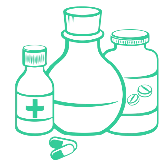
The department of General Medicine at North City Hospital is central to the clinical care of complex patients. Our medical staff specialize in the diagnosis and treatment of a broad range of diseases involving all organ systems, and are especially skilled in the management of patients with complex medical needs.
In our general medicine unit, treatment is provided through the expertise of the medical team and the multidisciplinary team. The team is comprised of a staff physician, the referred physician and the team of junior doctors. Patients are seen on a daily basis by one of the team members.
Prior to discharge, members of your health care team will discuss discharge plans with you and provide information on follow-up appointments, medications and other instructions
What is Diabetes?
Diabetes is a chronic condition characterised by high blood glucose levels (hyperglycaemia). Diabetes occurs when the pancreas loses its ability to produce enough insulin (a type of hormone), or when the body does not respond to insulin action. When blood glucose (sugar) levels increase, after we eat, the pancreas secretes insulin to help body cells convert glucose into energy, or to store it.
In people with Diabetes, instead of the glucose being converted to energy, it remains in the blood, therefore leading to higher than normal blood glucose levels. People with Diabetes have an increased risk of developing cardiovascular (heart-related) diseases, because it is often associated with high blood pressure, high cholesterol levels and obesity.
There are three main types of Diabetes:
Causes of Diabetes
Type 1 Diabetes is caused by the absolute lack of insulin in the body, due to the destruction of the pancreatic cells responsible for insulin secretion. Type 1 Diabetes is the most common cause of childhood diabetes. People with this form of diabetes require daily insulin injection to survive.
Type 2 Diabetes is marked by decreased levels of insulin or the inability of the body to use insulin properly (known as insulin resistance). The onset of this form of diabetes is usually gradual with symptoms generally appearing after the age of 40. Various risk factors can lead to Type 2 Diabetes including lack of physical activity, unhealthy diet, and obesity. People with Type 2 Diabetes often have a family history of the disease.
Gestational diabetes occurs in 2-5% of pregnant women who were not previously diagnosed with diabetes. It usually disappears after giving birth, however it is a marker of increased risk of developing Type 2 Diabetes later in life.
Symptoms
The most common symptoms of diabetes are:
Treatment in General Medicine
Type 1 Diabetes treatment includes:
Type 2 Diabetes treatment mainly includes lifestyle changes to control blood glucose level:
Complications of Diabetes
Heart disease Heart disease is the leading cause of diabetes-related deaths. Adults with diabetes have heart disease death rates about two to four times as high as that of adults without diabetes.
Remark: *Diabetic ketoacidosis and hyperosmolar nonketotic coma are medical conditions that can result from very high glucose level and biochemical imbalance in uncontrolled diabetes.
Diabetic ketoacidosis (DKA) is a serious complication of type 1 diabetes and, much less commonly, of type 2 diabetes. DKA happens when your blood sugar is very high and acidic substances called ketones build up to dangerous levels in your body.
It’s less common in people with type 2 diabetes because insulin levels don’t usually drop so low; however, it can occur. DKA may be the first sign of type 1 diabetes, as people with this disease can’t make their own insulin.
Symptoms
DKA is a medical emergency. Call your local emergency services immediately if you think you are experiencing DKA.
If left untreated, DKA can lead to a coma or death. If you use insulin, make sure you discuss the risk of DKA with your healthcare team and have a plan in place. If you have type 1 diabetes, you should have a supply of home urine ketone tests. You can buy these in drug stores or online.
If you have type 1 diabetes and have a blood sugar reading of over 250 milligrams per deciliter (mg/dL) twice, you should test your urine for ketones. You should also test if you are sick or planning on exercising and your blood sugar is 250 mg/dL or higher.
Call your doctor if moderate or high levels of ketones are present. Always seek medical help if you suspect you are progressing to DKA.
Treatment in General Medicine
The treatment for DKA usually involves a combination of approaches to normalize blood sugar and insulin levels. Infection can increase the risk of DKA. If your DKA is a result of an infection or illness, your doctor will treat that as well, usually with antibiotics.
Fluid replacement
At the hospital, your physician will likely give you fluids. If possible, they can give them orally, but you may have to receive fluids through an IV. Fluid replacement helps treat dehydration, which can cause even higher blood sugar levels.
Insulin therapy
Insulin will likely be administered to you intravenously until your blood sugar level falls below 240 mg/dL. When your blood sugar level is within an acceptable range, your doctor will work with you to help you avoid DKA in the future.
Electrolyte replacement
When your insulin levels are too low, your body’s electrolytes can also become abnormally low. Electrolytes are electrically charged minerals that help your body, including the heart and nerves, function properly. Electrolyte replacement is also commonly done through an IV.
What is Hypertension (HTN or HT), also known as high blood pressure (HBP), is a long-term medical condition in which the blood pressure in the arteries is persistently elevated. Usually hypertension is defined as blood pressure above 140/90, and is considered severe if the pressure is above 180/120.High blood pressure typically does not cause symptoms. Over time, if untreated, it can cause health conditions, such as heart disease and stroke.
Causes
The causes of Hypertension are unknown in 95% of patients. In 5% of cases, some specific conditions can be responsible for the high blood pressure, such as kidney disease, atherosclerosis and hormonal imbalance. There are also several risk factors that may increase your chances of developing hypertension including diabetes, obesity as well as a strong family history of the disease.
Symptoms
Hypertension usually does not lead to any symptoms, but in the long term it can damage various organs and lead to the following:
Treatment in General Medicine
Your doctor will evaluate your condition and discuss with you the range of treatment options available. These include a combination of:
Antihypertensive medications may also be prescribed, and these need to be taken regularly and permanently.
What is Deep vein thrombosis?
Deep vein thrombosis (DVT) is a serious condition that occurs when a blood clot forms in a vein located deep inside your body. A blood clot is a clump of blood that’s turned to a solid state. Deep vein blood clots typically form in your thigh or lower leg, but they can also develop in other areas of your body This condition is serious because blood clots can loosen and lodge in the lungs.
Cause
DVT is caused by a blood clot. The clot blocks a vein, preventing blood from properly circulating in your body. Clotting may occur for several reasons. These include:
Symptoms
Symptoms of DVT only occur in about half of the people who have this condition. Common symptoms include:
People with an upper extremity DVT, or a blood clot in the arm, may also not experience symptoms. If they do, common symptoms include:
People may not find out that they have deep vein thrombosis until they’ve gone through emergency treatment for a pulmonary embolism (blood clot in the lung).
A pulmonary embolism can happen when a DVT clot has moved from the arm or leg into the lung. When an artery in the lung becomes blocked, it’s a life-threatening condition and requires emergency care.
Treatment in General Medicine
DVT is a serious medical condition. Tell your doctor right away if you think you’re experiencing symptoms of DVT or go to the closest emergency room. A healthcare provider can check out your symptoms.
DVT treatments focus on keeping the clot from growing. In addition, treatment may help prevent a pulmonary embolism and lower your risk of having more clots.
Medication
Your doctor might prescribe medications that thin your blood, like heparin, warfarin, enoxaparin, or fondaparinux. This makes it harder for your blood to clot. It also keeps existing clots as small as possible and decreases the chance that you’ll develop more clots.
If blood thinners don’t work, or if you have a severe case of DVT, your doctor might use thrombolytic drugs. People with upper extremity DVT may also benefit from this medication.
Thrombolytic drugs work by breaking up clots. You’ll receive these intravenously. Read more about these drugs and how they can help prevent and destroy blood clots.
Compression stockings
If you’re at high risk for DVT, wearing compression stockings can prevent swelling and might lower your chance of developing clots.
Compression stockings reach just below your knee or right above it. Your doctor may recommend you wear these every day.
Filters
You might need to have a filter put inside the large abdominal vein called the vena cava if you aren’t able to take blood thinners. This form of treatment helps prevent pulmonary embolisms by stopping clots from entering your lungs.
But filters do have risks. If they’re left in for too long, they can actually cause DVT. Filters should be used for a short-term period, until the risk of thromboembolism is reduced and blood thinning medications can be used.
DVT surgery
Your doctor may suggest surgery to remove a DVT clot in your arm or leg. This is typically only recommended in the case of very large blood clots or clots that are causing serious issues, like tissue damage.
During a surgical thrombectomy, or surgery to remove a blood clot, your surgeon will make an incision into a blood vessel. They’ll locate and remove the clot. Then, they’ll repair the blood vessel and tissue.
In some cases, they may use a small inflating balloon to keep the blood vessel open while they remove the clot. When the clot is found and removed, the balloon is removed with it.
Surgery isn’t without risks, so many doctors will only use this treatment in severe cases. Risks include infection, damage to the blood vessel, and excess bleeding.
DVT exercise
The longer you sit, the greater your risk of developing a blood clot. If you have to be seated for long periods, there are exercises you can do while sitting to keep your legs moving and help circulate blood.
Knee pulls
Bend your leg, and raise your knee toward your chest. Wrap your knee with your arms for a greater stretch. Hold this position for several seconds, then do the same exercise on the other side. Repeat these stretches several times.
Foot pumps
Place your feet flat on the floor. Keeping the balls of your feet on the floor, raise your heels. Hold for a few seconds, then lower the heels. Raise the balls of your feet off the floor, keeping your heels in place. Hold for a few seconds, then lower the balls of your feet.
Repeat these pumps several times.
Ankle circles
Lift both feet off the floor. Draw circles with your toes in one direction for a few seconds. Switch directions, and draw circles for a few seconds. Repeat this exercise several times.
DVT home remedies
Once a DVT blood clot is diagnosed, your doctor will likely prescribe a medication to help thin the blood or break up the clot. You can combine the prescribed medication with the following home remedies to prevent other complications and reduce the risk of future blood clots.
Move more
Take walks daily to improve blood flow. Shorter, frequent walks are better than one longer walk.
Keep your leg or arm elevated
This is especially important for legs. Blood can pool if your feet are on the ground all day. Use a stool or chair to keep your legs elevated and close to level with your hips.
What is Electrolyte Imbalance?
Electrolyte imbalance is commonly caused by loss of body fluids through prolonged vomiting, diarrhea, sweating or high fever. Examples of electrolytes are sodium, chloride, magnesium, potassium and calcium. Electrolyte imbalance causes a variety of symptoms that can be severe.
Types of electrolyte disorders
Elevated levels of an electrolyte are indicated with the prefix “hyper-.” Depleted levels of an electrolyte are indicated with “hypo-.”
Conditions caused by electrolyte level imbalances include:
Potassium
Potassium is particularly important for regulating heart function. It also helps maintain healthy nerves and muscles.
Hyperkalemia may develop due to high levels of potassium. This condition can be fatal if left undiagnosed and untreated. It’s typically triggered by:
Hypokalemia occurs when potassium levels are too low. This often happens as a result of:
Sodium
Sodium is necessary for the body to maintain fluid balance and is critical for normal body function. It also helps to regulate nerve function and muscle contraction.
Hypernatremia occurs when there’s too much sodium in the blood. Abnormally high levels of sodium may be caused by:
Hyponatremia develops when there’s too little sodium. Common causes of low sodium levels include:
Calcium
Calcium is a vital mineral that your body uses to stabilize blood pressure and control skeletal muscle contraction. It’s also used to build strong bones and teeth.
Hypercalcemia occurs when you have too much calcium in the blood. This is usually caused by:
Hypocalcemia occurs due to a lack of adequate calcium in the bloodstream. Causes can include:
Treating electrolyte disorders in General Medicine
Treatment varies depending on the type of electrolyte disorder and on the underlying condition that’s causing it.
In general, certain treatments are used to restore the proper balance of minerals in the body. These include:
Intravenous (IV) fluids
Intravenous (IV) fluids, typically sodium chloride, can help rehydrate the body. This treatment is commonly used in cases of dehydration resulting from vomiting or diarrhea. Electrolyte supplements can be added to IV fluids to correct deficiencies.
Certain IV medications
IV medications can help your body restore electrolyte balance quickly. They can also protect you from negative effects while you’re being treated by another method.
The medication you receive will depend on the electrolyte disorder you have. Medications that may be administered include calcium gluconate, magnesium chloride, and potassium chloride.
Oral medications and supplements
Oral medications and supplements are often used to correct chronic mineral abnormalities in your body. This is more common in if you’ve been diagnosed with ongoing kidney disease.
Depending on your electrolyte disorder, you may receive medications or supplements such as:
They can help replace depleted electrolytes on a short- or long-term basis, depending on the underlying cause of your disorder. Once the imbalance has been corrected, your doctor will treat the underlying cause.
Although some of the supplements can be purchased over the counter, most people with electrolyte disorders get a prescription for supplements from their doctor.
Hemodialysis
Hemodialysis is a type of dialysis that uses a machine to remove waste from your blood.
Hemodialysis can be used when an electrolyte disorder is caused by sudden kidney damage and other treatments aren’t working. Your doctor may also decide on hemodialysis treatment if the electrolyte problem has become life-threatening.
What is Meningitis?
Meningitis is an inflammation (swelling) of the protective membranes covering the brain and spinal cord. A bacterial/viral or fungal infection of the fluid surrounding the brain and spinal cord usually causes the swelling.
Symptoms
In adults, viral meningitis may cause:
Bacterial meningitis symptoms
Bacterial meningitis symptoms develop suddenly. They may include:
Seek immediate medical attention if you experience these symptoms. Bacterial and viral meningitis can be deadly. There’s no way to know if you have bacterial or viral meningitis just by judging how you feel. Your doctor will need to perform various tests to determine which type you have.
Fungal meningitis symptoms
Symptoms of fungal meningitis resemble the other types of this infection. These may include:
Types and Causes of Meningitis
Each type of meningitis has a slightly different cause, but each ultimately acts in the same way: A bacterium, fungus, virus, or parasite spreads through the bloodstream until it reaches the brain, or spinal cord. There, it sets up in the lining or fluids around these vital body parts and starts developing into a more advanced infection.
Non-infectious meningitis is the result of a physical injury or other condition; it doesn’t involve an infection.
Viral meningitis is the most common type of meningitis. Viruses in the Enterovirus category cause 85 percent of cases. They include:
Viruses in the Enterovirus category cause about 10 to 15 million infectionsTrusted Source per year, but only a small percentage of people who get infected will develop meningitis.
Other viruses can cause meningitis. These include:
Viral meningitis typically goes away without treatment. However, some causes do need to be treated.
Bacterial meningitis
Bacterial meningitis is contagious and caused by infection from certain bacteria. It’s fatal if left untreated. Between 5 to 40 percentTrusted Source of children and 20 to 50 percentTrusted Source of adults with this condition die. This is true even with proper treatment.
The most common types of bacteria that cause bacterial meningitis are:
Fungal meningitis
Fungal meningitis is a rare type of meningitis. It’s caused by a fungus that infects your body and then spreads from your bloodstream to your brain or spinal cord.
People with a weakened immune system are more likely to develop fungal meningitis. This includes people with cancer or HIV.
The most common funguses related to fungal meningitis include:
Parasitic meningitis
This type of meningitis is less common than viral or bacterial meningitis, and it’s caused by parasites that are found in dirt, feces, and on some animals and food, like snails, raw fish, poultry, or produce.
One type of parasitic meningitis is rarer than others. It’s called eosinophilic meningitis (EM). Three main parasites are responsible for EM. These include:
Parasitic meningitis is not passed from person to person. Instead, these parasites infect an animal or hide out on food that a human then eats. If the parasite or parasite eggs are infectious when they’re ingested, an infection may occur.
One very rare type of parasitic meningitis, amebic meningitis, is a life-threatening type of infection. This type is caused when one of several types of ameba enters the body through the nose while you swim in contaminated lakes, rivers, or ponds. The parasite can destroy brain tissue and may eventually cause hallucinations, seizures, and other serious symptoms. The most commonly recognized species is Naegleria fowleri.
Non-infectious meningitis
Non-infectious meningitis is not an infection. Instead, it is a type of meningitis that’s caused by other medical conditions or treatments. These include:
Prevention in General Medicine
There is a vaccine for several types of bacterial meningitis. Meningococcal meningitis, caused by Neisseria meningitidis, is one version for which vaccines are available. While viral meningitis is more common, bacterial meningitis can be more dangerous if it’s not diagnosed and treated quickly.
For that reason, the two primary vaccines for meningitis are for bacterial causes. The first vaccine, the meningococcal conjugate vaccine, features a vaccine that targets four of the most common types of bacterial serotypes. It lasts longer and offers greater protection, especially if you maintain booster shots.
The second vaccine, MenB, targets one specific strain, and its protection window is much shorter. Only certain populations are recommended to get this vaccine
Who should be vaccinated against meningococcal meningitis?
These five groups are considered at risk and should get a meningitis vaccine:
Teenagers should protect themselves by getting a meningitis vaccine.
Treatment in General Medicine
Your treatment is determined by the cause of your meningitis.
Bacterial meningitis requires immediate hospitalization. Early diagnosis and treatment will prevent brain damage and death. Bacterial meningitis is treated with intravenous antibiotics. There’s no specific antibiotic for bacterial meningitis. It depends on the bacteria involved.
Fungal meningitis is treated with antifungal agents.
Parasitic meningitis may either involve treating just the symptoms or attempting to treat the infection directly. Depending on the cause, this type may get better without antibiotic treatment. If it worsens, however, your doctor may try to treat the infection itself.
Viral meningitis may resolve on its own, but some causes of viral meningitis will be treated with intravenous antiviral medications.
Complications
These complications are typically associated with meningitis:
What is Congestive Heart Failure?
Congestive heart failure (CHF) is a chronic progressive condition that affects the pumping power of your heart muscles. While often referred to simply as “heart failure,” CHF specifically refers to the stage in which heart muscle is too weak or when another health problem prevents it from circulating blood efficiently.
The most common type of HF is left-sided HF. The left side of the heart must work harder to move the same volume of blood around the body. This may cause a fluid buildup in the lungs and make breathing difficult as it progresses.
These fluids give congestive heart failure its name.
There are two kinds of left-sided HF:
Right-sided HF is less common. It occurs when the right ventricle cannot pump blood to the lungs. This can lead to blood backing up in the blood vessels, which may cause fluid retention in the lower legs and arms, abdomen, and other organs.
A person can have left-sided and right-sided HF at the same time. However, HF usually begins on the left side and can affect the right side if a person does not receive effective treatment.
Prevention
Lifestyle strategies can reduce the risk of developing HF and can also slow its progress.
To prevent or slow the progression of HF, people should take the following steps:
People who already have HF should take the following steps to prevent further progression:
Without treatment, HF can be fatal. Even with adequate treatment, HF may get worse over time, triggering dysfunction of other organs throughout the body.
Causes
HF is more likely to occur in people with other conditions or lifestyle factors that weaken the heart.
Risk factors for HF include:
Symptoms
People with a history of cardiovascular health issues or several risk factors for HF should seek immediate care if they experience symptoms of HF.
The most common symptoms of HF are:
Diagnosis
A doctor or cardiologist will perform a physical exam. This involves listening to the heart, checking for fluid retention, and looking at the veins in the neck to see if there is extra fluid present in the heart. They may order other diagnostic tests, including:
Treatment in General Medicine
Several types of medication can reduce the health impact of heart failure.
Different medications can help symptoms of and prognosis in HF. These include:
People with advanced HF might need more intensive treatment. Medical procedures that may help include the following:
Implantable devices
People with advanced HF might need more intensive treatment. A surgeon might implant a medical device, such as:
Other procedures
A doctor may recommend some other procedures for treating HF, including:
What Is Pneumonia?
It is an infection of the lungs with a range of possible causes. It can be a serious and life-threatening disease. It normally starts with a bacterial, viral, or fungal infection. The lungs become inflamed, and the tiny air sacs, or alveoli, inside the lungs fill up with fluid. Pneumonia blocks the normal exchange of gas inside the lungs, which leads to low levels of the oxygen in the blood and poor removal of carbon dioxide from the body. The severity of pneumonia ranges from mild to life threatening.
Causes
Pneumonia can affect anyone at any age and it can usually be triggered by a cold or a bout of flu.
Symptoms
Symptoms associated with pneumonia differ considerably depending on the cause and the health condition of the patient. These may include:
Treatment in General Medicine
Treatment of pneumonia depends on what triggered the infection. Your doctor will evaluate your condition and suggest the appropriate treatments for you. These may include:
What is Bronchial Asthma?
It is a medical condition which causes the airway path of the lungs to swell and narrow. Due to this swelling, the air path produces excess mucus making it hard to breathe, which results in coughing, short breath, and wheezing. The disease is chronic and interferes with daily working
Cause
No single cause has been identified for asthma. Instead, researchers believe that the breathing condition is caused by a variety of factors. These factors include:
Triggers of Bronchial asthma – Bronchial asthma triggers may include: Smoking and secondhand smoke. Infections such as colds, flu, or pneumonia. Allergens such as food, pollen, mold, dust mites, and pet dander.
Symptoms
Treatment in General Medicine
Treatment consists of self-care and bronchodilators
Asthma can usually be managed with rescue inhalers to treat symptoms (salbutamol) and controller inhalers that prevent symptoms (steroids). Severe cases may require longer-acting inhalers that keep the airways open (formoterol, salmeterol, tiotropium), as well as inhalant steroids.
Medications
Asthma home remedies
What is Hepatitis?
Hepatitis refers to the swelling of the liver. It can be caused by viral infections, chemicals, drug abuse, some medications and immune disorders. There are several forms of viral hepatitis including hepatitis A, B and C, which are caused by hepatitis A, B, and C viruses, respectively.
Each type of viral hepatitis is spread by different methods and needs different treatment.
Hepatitis A virus infection causes acute inflammation (swelling) of the liver and is a self-limiting disease with symptoms lasting for several weeks before the individual can recover completely. It leads to lifelong immunity.
Hepatitis B infection is the most common infection of the liver. The majority of infected individuals recover from acute hepatitis B infections and become immune to it. However some people can develop a long-term hepatitis B infection, which leads to serious complications including chronic hepatitis, liver cirrhosis (scarring of the liver), liver failure and liver cancer. Hepatitis B is endemic in Singapore and around 4% of the population are hepatitis B carriers.
Hepatitis C infection is responsible for the development of chronic liver disease worldwide. Most infected people can’t get rid of the virus, and thus the virus causes ongoing damage to the liver over the years. Similar to hepatitis B, hepatitis C can lead to chronic hepatitis, cirrhosis, liver failure and liver cancer.
Causes
Hepatitis A virus is transmitted through:
Hepatitis B virus is mainly found in the blood, and it can also be found in semen and vaginal secretions. Hepatitis B can be acquired through:
Hepatitis C virus is mainly found in the blood and is transmitted when the blood of an infected person enters the bloodstream of a susceptible person, like drug users sharing contaminated needles.
Symptoms
Some hepatitis patients are asymptomatic. However, the general symptoms may include any of the following:
Treatment in General Medicine
Hepatitis A
There is no specific treatment for hepatitis A but there are treatment measures that help improve your condition:
Hepatitis B
Treatment of hepatitis B depends on the symptoms and stage of your disease and includes:
Hepatitis C
Treatment of hepatitis C aims to delay its complications, and these include:
Complications
What is UTI?
A urinary tract infection (UTI) is an infection of any part of the urinary system. The urinary system consists of the kidneys, bladder, ureters and urethra. A urinary tract infection (UTI) is an infection of any part of the urinary system. UTIs usually affect first the bladder or the urethra, and if not treated they can then spread to the ureters and the kidneys.
The types of UTIs include:
Causes
Urine is usually sterile, which means it does not have any bacteria, viruses or fungi present.
A UTI can occur when a microorganism enters the urinary system through the urethra. Most infections are caused by Escherichia coli (E. coli), which is a digestive tract bacterium that lives in the colon, and spreads to the urethra from the anus.
Other microorganisms, including chlamydia and mycoplasma, can cause UTIs in men and women, but these UTIs are usually restricted to the urethra and the reproductive system.
Other causes that increase the risk of UTIs include:
Symptoms
UTI symptoms vary depending on the type of the infection and a person’s age. Some may not have any symptoms at all. When symptoms are present, they can include any of the following:
Treatment in General Medicine
| Name | Credentials |
| Prof. (Doctor) S. K. CHAKRABORTY | MBBS, DBMS, MD (CAL) |
| Prof. (Doctor) UDAS CH GHOSH | MBBS, MD (MED), DNB (MED), DNB (CHEST), DTCD (CAL) |
| Dr. ABHIJIT PAUL | MBBS, DTM & H, MD (CAL) |
| Dr. DEBABRATA MITRA | MBBS, DTM & H, MD, FIACM |
| Dr. ANUPAM CHAKRABORTY | MBBS, MD (MED) |
| Dr. GAUTAM SARKAR | MBBS, MD |
| Dr. AFIFUR RAHAMAN | MBBS, MD, MRCP, DNB |
| Dr. ALOK KUMAR GHOSH | MBBS, MD (CAL) |
| Dr. MUKUL KUMAR SAHA | MBBS, MD, DNB, MACP (USA) |
| Dr. S K MUKHARJEE | MBBS, MNAMS (AIIMS), MIPHA (INDIA) |
| Dr. P. S. MUKHERJEE | MBBS (B’LORE), MRCP (UK), FRCP (LOND), FACP, FASN (USA) |
| Dr. CHAYAN ROY | MBBS, DTM & H |
| Dr. ANIRUDDHA GHOSH | MBBS, A.C.D.M. (RCGP) |
| Dr. SAYAN GUPTA | MBBS, (HONS), MRCP (UK) LONDON, MRCP (IRELAND-DUBLIN) |
| Dr. RAKTIMAVA SARKAR | MBBS, MD |
| Dr. AMITABHA PAL | MBBS, MD |
| Dr. DEBAJYOTI MAJUMDAR | MBBS, MD, CCEBDM |
A positron emission tomography (PET) scan is an imaging test that allows your doctor to check for diseases in your brain. The scan uses a special dye containing radioactive tracers. … When detected by a PET scanner, the tracers help your doctor to see blood flow in the brain, to demonstrate areas of activity. This test is done at our Diagnostic Division.
Machine Used in General Medicine: Siemens mCT 128 with Ultra HD Technology
Positron emission tomography (PET) viability imaging is used to assess how much heart muscle has been damaged by a heart attack or heart disease. This test is used to determine whether a patient may need angiography, cardiac bypass surgery, heart transplant or other procedures.
Preparation – This test is done at our diagnostic branch .For details please contact our PET department
Machine Used in General Medicine- Siemens Biograph mCTx
Whole body PET CT in General Medicine is useful for detecting cancer and for:
In many cases of PUO like Extra Pulmonary TB, Sarcoidosis, Aortoarteritis, Lymphoma, etc., Whole Body PET CT can diagnose the disease.
A myocardial perfusion SPECT (Single Photon Emission Computed Tomography) study, also called a cardiac stress-rest test, is used to evaluate the heart’s blood supply.
Preparation – This test is done at our diagnostic branch .For details please contact our SPECT department
Machine Used in General Medicine – Siemens Symbia T
The MRI scan is an imaging test that allows physicians to assess a patient’s brain anatomy and investigate an anatomical cause of the patient’s Headache/Weakness,/Seizure /Blurring Of Vision etc. This test is done at our Diagnostic Division.
Machine Used in General Medicine – MRI Signa HDXT 1.5T
While magnetic resonance imaging (MRI) identifies the anatomical location of a tumor, MR spectroscopy compares the chemical composition of normal brain tissue with abnormal tumor tissue. This test can also be used to detect tissue changes in stroke and epilepsy. This test in General Medicine is done at our Diagnostic Division.
MRI of the heart lets your doctor see if your heart is damaged from a heart attack, or if there is lack of blood flow to the heart muscle because of narrowed or blocked arteries. MRI of the brain is used to diagnose stroke, aneurysm and other brain abnormalities. Viability assessment by MRI is a nonstress examination that provides high-resolution detail, including functional assessment of the left ventricle in approximately 30 minutes. Assessment of myocardial viability is performed using 5- to 20-minute delayed, gadolinium-enhanced MRI.
Preparation – This test is done at our diagnostic division.Nothing much preparation is needed. Though for details please contact our MRI department
Machine Used in General Medicine – GE Signa HdXT(1.5T)
Coronary computed tomography angiography (CCTA) uses an injection of iodine-containing contrast material and CT scanning to examine the arteries that supply blood to the heart and determine whether they have been narrowed. The images generated during a CT scan can be reformatted to create three-dimensional (3D) images that may be viewed on a monitor, printed on film or by a 3D printer, or transferred to electronic media.
Preparation – This test is done at our diagnostic division.4-6 hours empty stomach is needed. Though plain water can be taken.Urea, Creatinine, previous ECG, TMT, ECHOCARDIOGRAPHY, Prescriptions are needed.
Machine Used in General Medicine – Siemens Biograph mCTx
CT scans Of Brain can provide more detailed information about brain tissue and brain structures than standard X-rays of the head, thus providing more data related to injuries and/or diseases of the Brain. This test is done at our Diagnostic Division.
Machine Used in General Medicine – GE Bright Speed Elite CT
An electroencephalogram (EEG) is a noninvasive test that records electrical patterns in your brain. The test is used to help diagnose conditions such as seizures, epilepsy, head injuries, dizziness, headaches, brain tumors and sleeping problems. It can also be used to confirm brain death.
EMG results are often necessary to help diagnose or rule out a number of conditions such as: Muscle disorders, such as muscular dystrophy or polymyositis. Diseases affecting the connection between the nerve and the muscle, such as myasthenia gravis. This test is done at our Diagnostic Division.
A nerve conduction velocity (NCV) test — also called a nerve conduction study (NCS) — measures how fast an electrical impulse moves through your nerve. NCV can identify nerve damage. This test in General Medicine is done at our Diagnostic Division.
An ECG records the electrical activity of the heart. The ECG test is painless and harmless. An ECG may be part of a routine physical exam or it may be used as a test for heart disease like Diagnosing poor blood flow to the heart muscle (ischemia) or diagnosing a heart attack. Or evaluate certain abnormalities of your heart, such as an enlarged heart.
Preparation – No Preparation is needed. For details please contact our Cardioogy department
Machine used in General Medicine – GE MAC 600
It is also known as an exercise electrocardiogram, It lets your doctor know how your heart responds to being pushed. You’ll walk on a treadmill with the patient connected to an electrocardiogram (ECG).
Preparation – Consult your physician about going off beta blockers for 48 hours and calcium channel blockers 24 hours before your exam. Do not eat or drink for three hours before your appointment. For details please contact our Cardiology department.
Machine Used in General Medicine – GE Cardiosoft.
A Holter monitor is a battery-operated portable device that measures and tape records your heart’s activity (ECG) continuously for 24 to 48 hours or longer depending on the type of monitoring used. The device is the size of a small camera. It has wires with electrodes that attach to your skin.
Preparation – Nothing much preparation is needed. Though for details please contact our Cardiology department
An echocardiogram checks how your heart’s chambers and valves are pumping blood through your heart. An echocardiogram uses electrodes to check your heart rhythm and ultrasound technology to see how blood moves through your heart. An echocardiogram can help your doctor diagnose heart conditions.
2-D Doppler Echocardiogram or a echocardiogram with Colour Doppler measures the speed and direction of the blood flow within the heart. It screens the four valves for leaks and other abnormalities. By assigning color to the direction of blood flow, (Color Flow Mapping), large areas of blood flow may be studied.
Preparation – Nothing much preparation is needed. Though for details please contact our Cardiology department.
Machine Used in General Medicine – Philips Affinity 70
Ambulatory blood pressure in General Medicine monitoring allows many blood pressure (BP) readings to be recorded over a 24-hour period, whether the patient is awake or asleep.
Preparation – Nothing much preparation is needed. Though for details please contact our Cardioogy department.
In case of
Please call

Please fill up the form below to speed up the patient’s admission process